Introduction
Arthropathy can be caused due to crystal deposits like monosodium monohydrate (MSUM gout) or calcium pyrophosphate dehydrate (CPPD pseudogout). Decreased uric acid excretion is the main cause of gout.
Clinical features
Acute synovitis, tophi, chronic destructive and deformity arthritis, nephrolithiasis, interstitial nephritis and hypertension.
Hyperuricemia defined by blood urate concentration more than 7 mg percent. Only few with hyperuricemia present with gout. Hence hyperuricemia may be an incidental event and may not lead to gout.
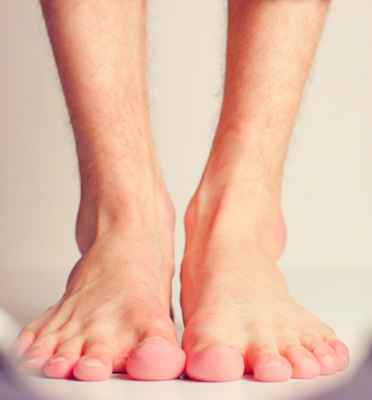
The big toe is the usual place for urate deposits. There may be other places affected like the instep, ankle, knee or joints of the hand. The surrounding area becomes inflammed. Many joints may be simultaneously affected.
Chronic gout with development of tophi may occur in prolonged uncontrolled hyperuricemia. Tophi look like firm swelling and may have inflammation. If the tophi become ulcerated, the urate crystals may come out in the form of white chalky matter.
Gout may result in kidney disease. It can give rise to uric acid stone or uric acid nephropathy
Investigation
Synovial fluid is turgid with decreased viscosity. It has increased neutrophils. Joint fluid can be seen under polarised light. Needle shaped urate deposits can be seen in synovial fluid and tophi.
Blood for hyperuricemia.
Predisposing factors
High blood urate
Thiazide diuretics, alcohol, starvation, dehydration,
High purine foods result in increased urate. Examples of high purine foods are red meat, liver, kidney and pulses.
Few blood disorders like leukaemia lymphoma, hemolytic anemia.